By Tony M. Woodward, DVM, AVDC
We continue describing the five basic veterinary dental services that all general practitioners should be able to provide for their patients. Previously we covered dental radiology. This article will discuss the rationale and veterinary dental equipment used for surgical extractions.
Historically, dental extractions have been viewed as necessary mainly when excessive tooth mobility is observed. Pet teeth exhibiting a high grade of mobility are easily extracted using dental elevators and forceps, in some cases falling out as soon as calculus is removed. For many general practitioners, if a tooth does not show mobility, it is left in regardless of what pathology is present. Because extractions on highly mobile teeth are easily accomplished, they have frequently been left to the veterinary technicians to perform. This attitude can lead to undercharging or not charging for extractions (“it only took the technician a few minutes”) and client expectations that extractions are free and are included with a dental cleaning procedure. It should be noted that extractions are considered a surgical procedure, and as such are illegal for technicians to perform in most states. This creates a legal liability for the supervising veterinarian who directs their technicians to perform this procedure.
Surgical extractions in pets involve the elevation of gingival flaps to provide adequate exposure, sectioning of multi-rooted teeth, cortical bone removal using a high-speed drill, luxation of the tooth segments with veterinary dental elevators, smoothing of the alveolar bone, and closure of the extraction site using muco-gingival flaps. When a clinician begins to aggressively look for and treat dental disease, they quickly realize that most needed extractions are on teeth with no mobility, and are best accomplished using a surgical technique. Surgical extraction techniques serve to improve exposure, increase patient comfort due to refinement in technique, decrease the incidence of complications such as root end fracture, and speed post-operative healing. For the above reasons, the author prefers to perform almost all extractions using a “surgical “technique. Some clinicians have preferred to leave extraction sites open, stating the need to allow drainage. Drainage is simply not required. In humans, extraction sites are not routinely closed. This is due to cosmetic considerations and the fact that human patient can be relied upon to keep extraction sites clean.
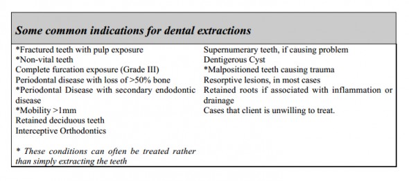
The indications for dental extractions include a wide variety of pathological conditions. A brief list is included below. This list should be tempered with the knowledge that many of these conditions can be treated. Whether to treat or extract a tooth depends on many factors. Some clients prefer to maintain all dentition if the prognosis is reasonably good for success. Certainly, the option of treatment vs. extraction should be offered when applicable, if only for liability reasons. Some teeth are more important for function such as the carnassial teeth (upper fourth premolar and lower first molars), and as such deserve consideration for treatment rather than extraction. Extraction of some teeth involves a large amount of bone removal, which can be painful and damage or weaken surrounding structures. An example would be a fractured mandible which occurred secondary to extraction of a lower canine tooth. For reasons previously stated, clients often think of an extraction as a cheap, fast treatment option for diseased dentition. Frequently, extraction of a diseased tooth is more painful, takes longer, and decreases function more than treatment of that same tooth. Teeth that particularly deserve treatment consideration
include the canine teeth and the carnassial teeth. In certain breeds, all of the anterior dentition can be important to show success. The astute clinician presents all options for treatment, along with the pros and cons of each option. Let the client decide which option they prefer.
A frequently misunderstood area of veterinary dentistry involves feline extractions. I have heard many people state that when extracting teeth in feline patients, one has to simply amputate the crowns, leaving the roots to magically reabsorb. This is a barbaric practice. How would you feel if your dentist amputated your crown, leaving an exposed amputated root fragment and nerve tissue. Would you feel better knowing that in a few years the root would be reabsorbed and the area healed over? Many of my feline patients have chronic draining tracts with root fragments left under the gum line. After treatment, owners notice these patients acting much better. This misunderstanding dates back to a 1995 article on intentional root retention. This article stated that this technique can be used if:
- There is radiographic evidence of bony replacement resorption.
- There is no evidence of endodontic disease.
- There is no evidence or periodontal disease
- There is no ulcero-proliferative disease (Stomatitis)
- The dental tissues are amputated 2-3 mm below the level of the alveolar bone.
- The extraction site is sutured closed.
Unfortunately, many people forget the six caveats stated above, and simply choose to remember that in cats, you only need to amputate the crowns. It is always safer and better for the patient to remove all remnants of the tooth when performing extractions.
Dental Instrumentation
Like all other surgical techniques, having the proper equipment makes the procedure go more smoothly and facilitates a more professional result. The following list includes the instruments and materials that the author finds useful for surgical extractions.
- High-speed delivery unit with integrated water coolant
- Burs for high-speed handpiece: 1/2, 1. 2, 4, 700, 701,701L, cylindrical diamond, conical white stone
- Scalpel blades: #11, #15, #15C
- Periosteal elevators: Molt #2, Molt #4, and Freer
- Dental elevators: Winged Elevators #1-4; Luxators in various sizes
- Extraction forceps: Small rongeurs work well
- Small Alveolar Curette
- Tissue Forceps: Brown-Adson 7X7 tooth
- Olsen-Hegar Needle holders, 5 1/2 inch
- Scissors: Dean
- Excellent lighting
- Dental Radiographs (mandatory)
- Gel-foam for hemostasis
- Consil synthetic allograft for bone augmentation
- Suture: Dexon, Vicryl, Monocryl, or Chromic Gut in 3-0 to 5-0 sizes, with reverse cutting or tapered needle. My personal preference is to use 5-0 Dexon for almost all extractions.
- Root tip picks for retrieval of fractured root tips
Many of these specific extraction instruments are carried in the Woodward Extraction Kit
produced by Dentalaire. I can perform 98% of all my extraction work utilizing this instrument set.
There are several good dental delivery units available on the market. I own six of the Dentalaire units which we use to teach classes. These have proven to be very dependable units under a hard work load. I recommend that you look at Dentalaire’s Ultimate unit, which includes a high quality piezoelectric scaler and fiber-optic lighting built into the high speed handpiece.
The combination low speed motors/scaler units are very adequate for cleaning procedures, but function poorly for extractions. The low speed micro-motor handpieces only turn at a maximum speed of 35-40,000 rpm, compared to 400,000 rpm for a compressor driven high speed handpiece. The micro-motor handpieces lack an integrated water coolant, and frequently burn bone and bog down as the operator attempts to section teeth and remove bone. If you have suffered through extractions using a micro-motor, you will be pleasantly surprised at how much easier procedure go with a high-speed unit.

