By Tony M. Woodward, DVM, AVDC
We continue describing the five basic veterinary dental services that all general practitioners should be able to provide for their patients. Earlier we covered the indications and equipment needed for surgical pet teeth extractions. This article will cover the specific techniques employed when performing this procedure.
General Principles of Surgical Extractions
There exist certain principles regarding surgical extraction that hold true regardless of the
specific tooth involved.
- When extracting a tooth, one should always endeavor to extract the roots in their entirety. Retained roots frequently are associated with draining tracts, which can be uncomfortable for the patient. When radiographs show ankylosis (fusion) of the root to the surrounding bone, root fragments are sometimes left behind intentionally. In these cases, the additional trauma for extracting the ankylosed roots is deemed to outweigh the risk of the root not being reabsorbed over time.
- Tissue handling should always be gentle to promote tissue vitality. Particular care should be used when handling delicate gingival flaps.
- When using dental elevators, one should always have respect for the forces generated. Due to the large amount of leverage, dental elevators can generate tremendous forces during normal use, and have potential to cause iatrogenic damage. Mandibular fractures can easily occur when extracting lower carnassial teeth and mandibular canines, especially if pre-existing periodontal disease has already weakened the bone.
- Dental instruments such as elevators and root tip picks can easily “spear” vital structures.They should always be used with a forefinger placed near the tip to serve as a “stop” to limit how far the instrument penetrates in the event of a slippage. Structures that can be injured include the Mandibular Artery, Vein, and Nerve which run in the Mandibular Canal, the Infraorbital Artery, Vein, and Nerve which run in the Infraorbital Canal above the upper carnassial tooth (PM4) and molars, the globe, the Maxillary Artery, and the Major Palatine Vessels. These structures will be covered in more detail later.
- Always give yourself adequate exposure to extract a tooth. Creating and later suturing a muco-gingival flap creates extra work, but it greatly simplifies the extraction process and provides a net savings of time. The author utilizes flaps for almost all extractions.
- Dental radiographs should always be used prior to extracting teeth. Frequently you will find information that will modify your extraction procedure. Additional radiographs during the procedure will help keep you on track, and can provide valuable information about retained roots.
General Technique for Surgical Extractions:
- Radiograph to determine the anatomy involved.
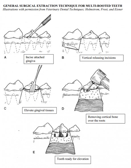
- Incise the epithelial attachment in the gingival sulcus with a #11 or # 15c scalpel blade.
- Elevate the attached gingiva over the extraction site using a periosteal elevator. Generally, the gingiva should be elevated at least to the level of the muco-gingival line. To allow additional exposure and lengthening of the flap for closure you can add vertical releasing incisions. These incisions should start between the tooth to be extracted and the adjacent healthy tooth, leaving a collar of attached gingiva untouched around the healthy tooth. These incisions should diverge from one another as they proceed apically, and should extend through the muco-gingival line into the mucosa. The flap is then termed a muco-gingival flap. The divergence of the incisions creates a broad based flap with a good blood supply, and adequate width for tension-free closure. After the attached gingiva of the flap is elevated, further elevation into the mucosa allows for lengthening of the flap. Incision of the periosteal layer on the underside of the flap provides for even more length, which is the key to a tension-free closure.
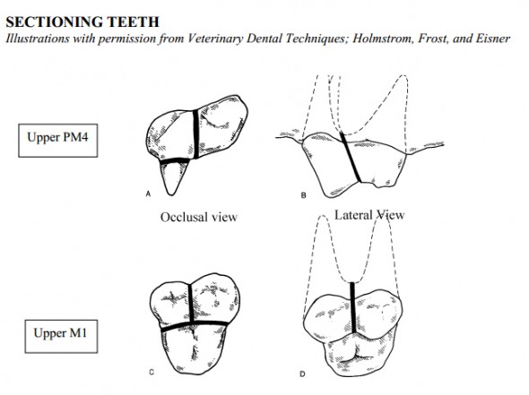
- Section the tooth if a multi-rooted tooth. This effectively transforms the multi-rooted tooth into several single-rooted teeth. This is particularly helpful when one realizes that multi-rooted teeth usually have divergent roots, which makes extraction very difficult unless the teeth are sectioned. The sectioning should be done through the furcation, taking care to section the entire crown. You can verify that the tooth is completely sectioned by placing a small instrument, such as a dental elevator, between the crown fragments. Gentlytwist or lever the instrument to separate the crown fragments. If the crown is completely sectioned, you will observe the crown fragments moving apart when gentle pressure is applied
- Remove the lateral (buccal or labial) cortical bone from the coronal 1/3 to 1/2 of the root. This allows much easier elevation and decreases the chance of root tip fracture during extraction. Removing a small amount of bone from the mesial and distal (cranial and caudal) root surfaces may allow for easier initial placement of the dental elevator. Removing this bone give the root a direction to luxate as you extract it. Teeth with some mobility might need little or no bone removal, while a lower first molar with a curved root tip might require more aggressive bone removal.
- Using a dental elevator, extract the roots. Place the edge of the elevator into the periodontal ligament space, and gently rotate and hold for 5-10 seconds to loosen the root. Repeat as you work your way around the root. The thin design of the luxators and winged elevators allow them to be placed into the ligament space with relative ease. You may also place the elevator between the crown fragments in a tooth that you have sectioned, taking care to not fracture the crown. Rotational forces are the primary forces used when using dental elevators.
- Remove the roots using an extraction forcep. Use forceps only after the tooth/root fragment is almost loose enough to remove with your fingers. You can gently rotate the fragment back and forth to help loosen it. If you force it, you will fracture roots frequently! Curved baby ronguers make ideal extraction forceps, and are small enough for use in both dogs and cats.
- Remove any fractured root fragments. This might involve further cortical bone removal, additional radiographs, the use of root tips picks, or other techniques. Take care not to force root tip fragments into the mandibular canal, maxillary recess, or nasal passages. You can use a cylindrical diamond bur to help make a small base of bone with the root visible in the center. You can then use a small round bur (#1/2) to outline the root and create a space for your dental elevator. Root tip picks are very thin and are useful when retrieving broken roots. They must be used with caution since it is easy to “spear” vital structures during their use.
- Curette and lavage the alveolus to remove loosed debris and infectious material.
- Smooth all sharp bony edges using rongeurs, bone rasps, or cylindrical diamond burs. Smoothing the bony contour prior to closure improves flap healing, increases patient comfort, and decreases tension on the flap.
- Suture the flap to cover the extraction site. The flap may need to be lengthened further by incising the periosteum or undermining the connective tissue. Place sutures 2-3mm apart, taking tissue bites of 2-3 mm. There should be no tension on the flap, and the suture line should ideally be placed over bone rather than over the alveolus. Appropriate suture is 3-0 to 5-0 size, using absorbable suture such as Dexon, Vicryl, Monocryl, or Chromic Gut.
- You may choose to place an osteopromotive material, such as Consil, into the extraction site to promote faster bone growth in the alveolus and help maintain the height of the alveolar ridge. The author finds this to be helpful in extraction sites of lower canine teeth and lower first molars, since extraction weakens the remaining mandibular structure significantly. These materials also assist in hemostasis. The use of an osteopromotive material might also be considered after extracting the upper canine tooth in cats, as this may help maintain the bulge of the alveolus and prevent “lip catch” of the upper lip on the lower canine tooth. Ensure that an oronasal fistula is not present prior to using an osteopromotive material in a maxillary extraction site. If a fistula is present, the material will be displaced into the nasal passages or maxillary recess.
- Pain control may be enhanced by placing a small amount of a local anesthetic agent such as Mepivicaine or Bupivicaine into the sutured extraction site (“splash block”). Do not exceed a total body dose of 2 mg/kg when utilizing these agents.
- Aftercare usually includes softening food for 10-14 days post-operatively, limiting access to hard chew toys, limiting rough play, Chlorhexidine oral rinse twice daily, and possibly antibiotics for 7-10 days. The author prefers to re-check all extraction sites 2-3 weeks post-operatively. This provides another opportunity to compliment the owner on the care they have provided for their pet.
Cautions for surgery around specific teeth
- Upper Canine: The infraorbital neurovascular bundle exits the foramen located above the third premolar, and courses cranially in the connective tissue. The thin cortical plate on the palatal (medial) aspect of the apical region of the root may be fractured if the root is levered palatally. Elevating the palatal (medial) aspect of the root should be done very cautiously to prevent the formation of an oro-nasal fistula. Improper flap design may also contribute to the formation of an oro-nasal fistula at this extraction site.
- Upper PM4 (Carnassial): The Infraorbital canal with it’s associated neurovascular bundle runs directly above this tooth, between the apices of the two mesial (cranial) roots, and above the apex of the distal root.
- Upper M1/M2: The Infraorbital canal runs above these teeth, and the Maxillary Artery runs in the retrobulbar space prior to entering the Infraorbital Canal. Additionally, the globe itself is easily damaged if an instrument slips during extraction.
- Lower Canine: This tooth makes up around 70-80% of the mandibular cross-section. Extraction of this tooth significantly weakens the mandible, predisposing toward iatrogenic fracture. The risk is increased in the presence of periodontal disease.
- Lower M1 (Carnassial): The roots of this tooth may extend to the ventral mandibular cortex, especially in small dogs. The roots may be locating lateral, medial, or within the mandibular canal. Root tips may be displaced into the mandibular canal, and the large mandibular neurovascular bundle is easily damage by overzealous elevation. The roots are frequently hooked or bulbous, which may increase the difficulty of extraction. Mandibular fractures are frequently associated with extraction of this tooth when affected by periodontal disease. Pre-extraction radiographs are strongly recommended to fully assess the anatomy and any pre-existing disease prior to extraction. Any perceived additional risks of extraction should be communicated to the owner prior to proceeding


